Deep Assessment of Neuroinflammation Using CNS Imaging and Analysis of CSF, Blood, Saliva Samples
Study AIM
To explore the hypothesis that low-grade neuroinflammation in the central nervous system can be part of the cardinal symptoms of ME/CFS.
LEAD INVESTIGATORS
Jonas Bergquist, MD, PhD
Wenzhong Xiao, PhD
Björn Bragée, MD
Updates and Potential
The analysis of cerebrospinal fluid and matching plasma samples are ongoing and manuscripts are in preparation.
Study protocol for MRI and PET imaging study was approved by the IRB in September 2024. Recruitment has been delayed due to the development and validation of a novel PET tracer.
STUDY HYPOTHESIS AND DESCRIPTION
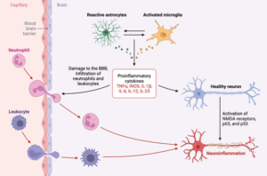
The overall aim is to explore the hypothesis that low-grade neuroinflammation in the central nervous system can be part of the cardinal symptoms of ME/CFS. Accumulating evidence suggests that ME/CFS patients can suffer from chronic neuroinflammation, the sustained activation of microglia and astrocytes, to strongly influence neurocognitive disturbances, headache, and fatigue and contribute to its progression. A major question is whether inhibition of the inflammatory response has the ability to reverse or slow down its symptoms. Furthermore, for many other brain diseases, such as neurodegenerative disorders, neuroinflammation is emerging as a cause, rather than a consequence, of the pathogenesis.
Neuroinflammation can possibly be monitored in cerebrospinal fluid (CSF) as a result of the inflammatory reaction itself or as secondary markers of cell degradation and impaired cell repair mechanisms. Our study will be the first to systematically measure CNS structural abnormalities in a large ME/CFS case-control study using CSF collection and in-situ measurement of intracranial pressure together with the study of microglia activation by the application of a novel diffusion-based MRI methodology. Added to this will be collected biospecimens to perform extensive state-of-the-art analyses with the ambition to identify diagnostic and prognostic biomarkers.
OBJECTIVES
Characterize neuroinflammation by CSF composition analyses, monitoring inflammatory markers, as well as markers of cell damage and impaired cell repair mechanisms.
Characterize the prevalence and severity of CNS structural abnormalities and microglia activation using structural and diffusion-based magnetic resonance imaging (MRI).
Characterize CSF pressure and flow using noninvasive and invasive techniques during lumbar puncture.
Identify biomarkers of the disease by extensive and detailed analyses of CSF and blood using proteomics and metabolomics.
Conduct and analyze special magnetic resonance imaging of neuroinflammatory involvement of activated microglia in patients with ME/CFS (and later Long COVID) for an available neuroinflammatory detection tool and to be used in outcome measures in clinical trials.
Microvesicles are a type of extracellular vesicle that is released from the cell membrane. Vesicles are small, fluid-filled sacs or vacuoles within the body. Sequencing is a technique used to determine the exact sequence of bases (A, C, G, and T) in a DNA (or viral) molecule.
NMR Metabolomics
NMR (Nuclear Magnetic Resonance) is an instrument/tool used to perform metabolic studies, metabolic profiling, and metabolomics in biofluids and tissues for more than 40 years using magnetic fields. There is a very close connection between metabolic measurements and NMR. This connection has flourished because of NMR’s many unique strengths for characterizing complex mixtures’ chemical composition.
Proteomics
The large-scale study of proteins, which are large, complex molecules that are required for structure, function, and regulation of the human body's tissues and organs.
Autoantibodies
Antibodies that react with self-molecules and that occur in healthy individuals and are referred to as natural antibodies or autoantibodies.
Extracellular DNA for viral reactivation and Mitochondrial DNA
exDNA (extracellular DNA), exDNA is often secreted actively and is used to perform several tasks, thereby offering an attractive target or tool for biotechnological, medical, environmental, and general microbiological applications. Viruses are intracellular parasites that rely to a significant extent on the host cell for replication. Viral reactivation occurs when an active replication of the viral genome results in a lytic (degradation of the cell) infection characterized by the release of new progeny (descendant) virus particles.
Immune cell profiling
The immune system has many important regulatory roles in disease development and progression. Given the emergence of effective immune therapies, reliable predictors of response are needed. Immune cell profiling determines response by evaluating immune cell populations from treated and untreated samples. For our purposes, we will evaluate the white blood cell response to the viral infection using a process called “Cytof.”
Leukocyte Genomics
A leukocyte is a colorless cell circulating in the blood and body fluids and is involved in counteracting foreign substances and disease. A genome is all genetic material of an organism. Genomics is a biology field focusing on the structure, function, evolution, mapping, and editing of genomes. Therefore, leukocyte genomics is the study of all genetic material of leukocytes.
Metabolomics
Metabolomics is a way to study metabolism – that is, through measuring amounts of the metabolites (small molecules) produced by our bodies as we convert food into energy and other molecules that our cells need to survive. Metabolomics technology is ‘large-scale,’ meaning that several thousand metabolites can be measured from a single sample of e.g., blood or urine.
Micro RNA
Cells use Micro RNA to control whether a particular gene is making too much, too little or the normal amount of its protein at a particular time.
 Characterize neuroinflammation by CSF composition analyses, monitoring inflammatory markers, as well as markers of cell damage and impaired cell repair mechanisms.